Science / Explained
SERIES: How GLP-1 WorksArticle 5 of 5
GLP-1 sends its fullness signal up the vagus nerve

The vagus nerve is one of the main communication routes between the digestive tract and the brain. GLP-1 works inside that larger gut-brain signaling system.

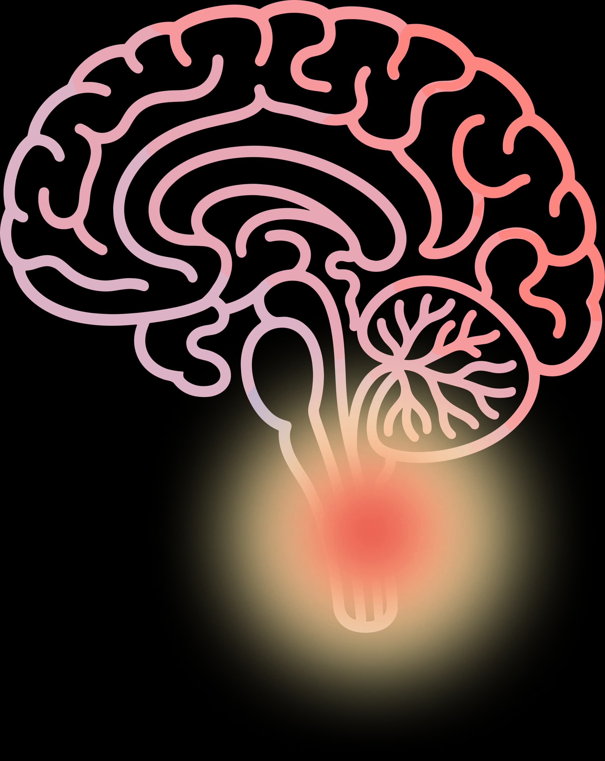
The vagus nerve connects the digestive tract to the brainstem and carries information upward from the gut. It is the longest nerve connecting the brain to the body, running from the brainstem through the neck and chest into the gut.

Signals related to stretch, nutrients, hormones, and internal state move through this route and help shape appetite, satiety, and reward. The traffic is not balanced. Roughly 80 percent of vagal signals flow upward, gut to brain. The brain sends some instructions down, but mostly it listens. It receives information about what the gut has eaten, what nutrients are present, and how digestion is proceeding.

GLP-1 works inside that system. After eating, the gut releases GLP-1 as part of the signaling response. The vagus nerve carries part of that signal directly to the brainstem. A second route runs through the bloodstream, reaching brain regions where the blood-brain barrier thins.

The vagus nerve is not the whole GLP-1 story. But it explains why digestion and brain response are linked so tightly. The gut is not waiting for the brain to ask questions. It is broadcasting constantly.

OneMoreThing

Not all vagus nerves transmit equally.Vagal tone measures how efficiently the nerve carries signals.Higher vagal tone correlates with better emotional regulation, lower resting inflammation, and stronger gut-brain communication.

Vagal tone varies significantly between individuals.This variation may partly explain why two patients on identical GLP-1 doses report entirely different experiences.A stronger cable carries a stronger signal.Researchers are now investigating whether measuring vagal tone could predict who responds best.The nerve is not the variable.Its efficiency is.

Previous
GLP-1 carries a signal from the gut to the brain
References3 sources

How to read these sources

This article uses primary sources and reviews to separate mechanism, human evidence, and context.

ReviewExpert synthesis
MechanismCell and pathway logic
Show 3 more source types
Official LabelRegulator documents
Human TrialStudies in people
Public UpdateNews or announcements
  1. Review

    Neurogastroenterology & Motility

    Wiley

    Neuroanatomy of extrinsic afferents supplying the gastrointestinal tract. Read source

    Used Here For

    Backing the anatomy of vagal and other nerve pathways carrying GLP-1-related gut signals.

    Good For

    A broad map of the nerves connecting the gut to the central nervous system.

    Not For

    Specific outcome data on any GLP-1 medicine.

    Neurogastroenterology & Motility, 16(Suppl 1)
  2. Review

    Journal of Clinical Investigation

    American Society for Clinical Investigation

    GLP-1 physiology and pharmacology along the gut-brain axis. Read source

    Used Here For

    Explaining how GLP-1 signals along the gut-brain axis, including via the vagus nerve.

    Good For

    A current synthesis of GLP-1 physiology and pharmacology research.

    Not For

    Patient-specific treatment or dosing guidance.

    Journal of Clinical Investigation, 156(3)
  3. Mechanism

    Journal of Clinical Investigation

    American Society for Clinical Investigation

    Neuroepithelial circuit formed by innervation of sensory enteroendocrine cells. Read source

    Used Here For

    Explaining the neuroepithelial circuit that links gut sensory cells to vagal afferent signaling.

    Good For

    Understanding the cellular wiring behind gut-to-brain signaling.

    Not For

    Clinical treatment guidance or patient-specific claims.

    Journal of Clinical Investigation, 125(2)